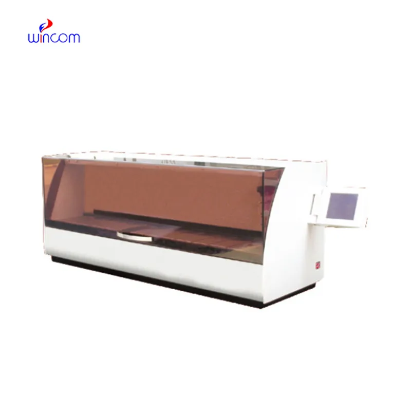
c18 hplc column enables labs to separate and analyze intricate mixtures with utmost precision. Through a seamless connection with current detectors, the method provides detailed profiling of both chemical and biological substances. The researchers and therapists trust c18 hplc column for the purposes of monitoring outcomes of experiments, method development, and cross-analyses accuracy. Its strength in dealing with various kinds of samples renders it an indispensable device in both the research and the clinical settings, thus improving reproducibility and backing up the struggling with more complex scientific and medical inquiries.
Hospital laboratories depend on c18 hplc column for identifying minute quantities of pharmaceuticals and therapeutic agents in difficult-to-analyze biological samples. Its use spans drug compliance testing, pharmacokinetics profiling, and tracking medications after surgery. The laboratory personnel can rely on it for exact measurement, thus increasing the efficiency of clinical treatment.
The instruments for c18 hplc column of the future will be equipped with separation methods in multiple dimensions and fully automated sample preparation. The detection of trace amounts of metabolites, drugs, and biomarkers will be so accurate that hospitals and clinical laboratories will be the first to reap the benefits. The applications of c18 hplc column in the future will greatly help in complex diagnostics, research studies, and laboratory efficiency.
Preventive maintenance is c18 hplc column that play a very important role in clinical and hospital laboratories. The routine performance of flushing columns, cleaning injector valves, and monitoring pressure stability extends the life of the system. The laboratory staff is required to keep records of maintenance activities, replace consumables in a timely manner, and use solvents that are compatible. All of these practices are essential for the instruments' performance retention, lifespan extension, and high-quality analytical results, both in patient sample testing and research.
c18 hplc column is a standard method in diagnostic laboratories of hospitals to keep an eye on patients’ biochemical and therapeutic figures. It quantifies drugs, hormones, and small molecules accurately. c18 hplc column speeds up the clinical decision-making processes of physicians and facilitates treatment modifications by supplying them with quick and precise results. It is used by hospital labs for basic patient testing, pharmacokinetic studies, and special analyses. The method’s high reproducibility makes certain that the outcomes are consistent, whereas its versatility allows for the support of many clinical applications. c18 hplc column has turned into an irreplaceable instrument in hospital diagnostics, which not only enhances patient management but also provides healthcare professionals with thorough molecular information.
Q: Do you need special training for HPLC operation? A: The answer is yes, training is a prerequisite to accurately and safely using pumps, columns, and detectors. Q: What type of maintenance does HPLC have? A: It requires cleaning, flushing, and inspection of all components as well as calibrating. Q: Is it possible to use HPLC in drug monitoring? A: Sure, it is a common practice in hospitals to monitor the levels of therapeutic drugs and also to identify metabolites in the samples taken from the patients. Q: What is the duration of analysis using HPLC in a typical case? A: The analysis time can range from a few minutes to more than an hour depending on the nature of the sample and the kind of column used. Q: Is HPLC a good choice for environmental testing? A: Yes, it can be used to find out the presence of pollutants, pesticides, and other harmful substances in water, soil, and air samples.
The centrifuge operates quietly and efficiently. It’s compact but surprisingly powerful, making it perfect for daily lab use.
I’ve used several microscopes before, but this one stands out for its sturdy design and smooth magnification control.
To protect the privacy of our buyers, only public service email domains like Gmail, Yahoo, and MSN will be displayed. Additionally, only a limited portion of the inquiry content will be shown.
Could you please provide more information about your microscope range? I’d like to know the magnif...
Hello, I’m interested in your centrifuge models for laboratory use. Could you please send me more ...
E-mail: [email protected]
Tel: +86-731-84176622
+86-731-84136655
Address: Rm.1507,Xinsancheng Plaza. No.58, Renmin Road(E),Changsha,Hunan,China

 af
af
 es
es
 ar
ar
 tr
tr
 sw
sw
 pt
pt
 th
th
 ur
ur
 bn
bn
 ne
ne
 vi
vi
 km
km
 lo
lo
 de
de
 ru
ru
 fi
fi
 nl
nl
 fa
fa
 fr
fr
 ko
ko