With video displayed through beamforming and noise-filtering technology, the fetal doppler machine is able to present an image that is very sharp and stable. Easy-to-use touch-screen controls help to streamline the process while rapid image rendering is guaranteed by the fast processing. Equipped for contemporary healthcare settings, the fetal doppler machine is capable of working with both 2D and Doppler imaging.

The fetal doppler machine has demonstrated its irreplaceable nature in prenatal screening, cardiovascular diagnostics, and overall health evaluations.The fetal doppler machine is a technique that evaluates organ function, reveals pathological changes, and supports medical education by providing live imaging demonstrations. The fetal doppler machine technology gives doctors the ability to perform accurate and instantaneous assessments in a variety of clinical situations.

Through continued innovations in digital technology, the fetal doppler machine can be expected to improve and extend its applications within preventive medicine and telemedicine. The next generation of such technologies will facilitate collaboration among experts in real-time using cloud-imaging solutions. The fetal doppler machine can also work within wearables that include biosensors.

In order to extend the service life of the fetal doppler machine, it is recommended that users refrain from applying much force during the process of connecting/disconnecting probes. Power cables should always remain dry. The fetal doppler machine needs diagnostic tests to ensure that it produces quality images.
The fetal doppler machine uses state-of-the-art ultrasound technology to deliver real-time imaging for diagnostic and monitoring purposes. It aids physicians in assessing organs, blood vessels, and soft tissue with unmatched clarity. The non-surgical device is an important tool for guiding medical procedures and making precise diagnoses. The fetal doppler machine combines portability with precision, rendering it extremely useful in routine exams as well as emergency applications.
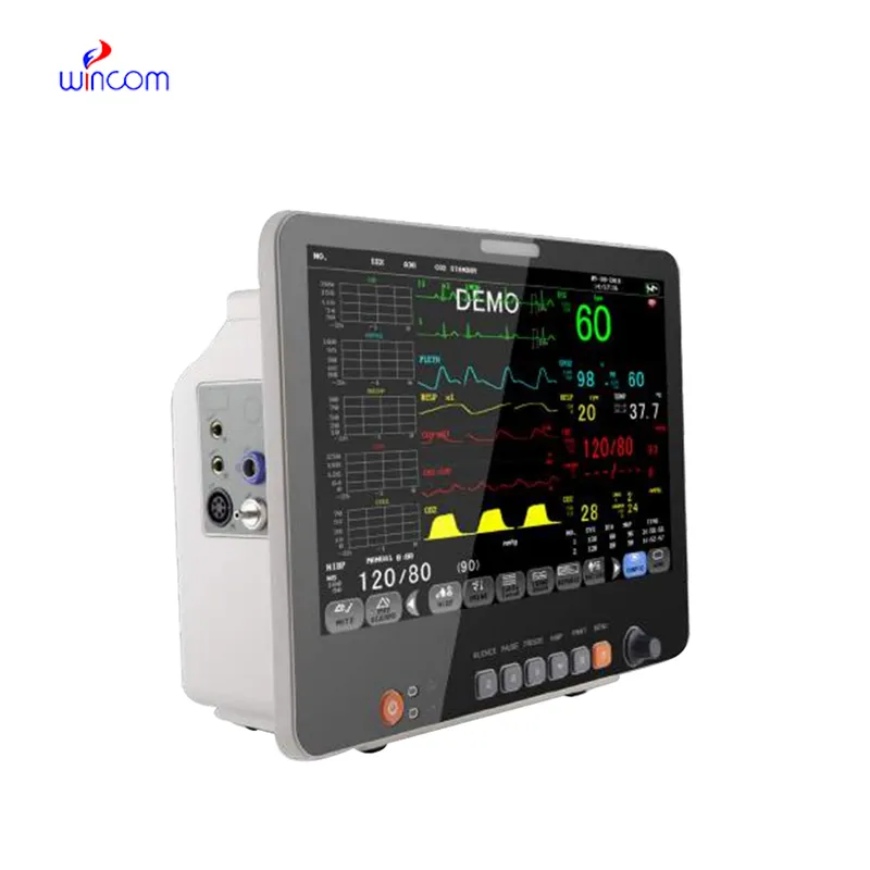
Q: What are the main maintenance requirements for the ultrasound scannert? A: Regular cleaning, proper probe handling, and scheduled inspections help maintain optimal performance. Q: How often should the ultrasound scannert be calibrated? A: Calibration frequency depends on usage levels, but periodic professional checks are recommended. Q: Is the ultrasound scannert suitable for pediatric use? A: Yes, it provides gentle, non-invasive imaging ideal for neonatal and pediatric diagnostics. Q: Does the ultrasound scannert support wireless connectivity? A: Many models include Wi-Fi or Bluetooth features for data sharing and device integration. Q: What materials are used in the ultrasound scannert construction? A: It is built with durable medical-grade components designed to withstand continuous clinical use.
I’ve used several microscopes before, but this one stands out for its sturdy design and smooth magnification control.
This x-ray machine is reliable and easy to operate. Our technicians appreciate how quickly it processes scans, saving valuable time during busy patient hours.
To protect the privacy of our buyers, only public service email domains like Gmail, Yahoo, and MSN will be displayed. Additionally, only a limited portion of the inquiry content will be shown.
Hello, I’m interested in your centrifuge models for laboratory use. Could you please send me more ...
I’m looking to purchase several microscopes for a research lab. Please let me know the price list ...
E-mail: [email protected]
Tel: +86-731-84176622
+86-731-84136655
Address: Rm.1507,Xinsancheng Plaza. No.58, Renmin Road(E),Changsha,Hunan,China

 af
af
 es
es
 ar
ar
 tr
tr
 sw
sw
 pt
pt
 th
th
 ur
ur
 bn
bn
 ne
ne
 vi
vi
 km
km
 lo
lo
 de
de
 ru
ru
 fi
fi
 nl
nl
 fa
fa
 fr
fr
 ko
ko