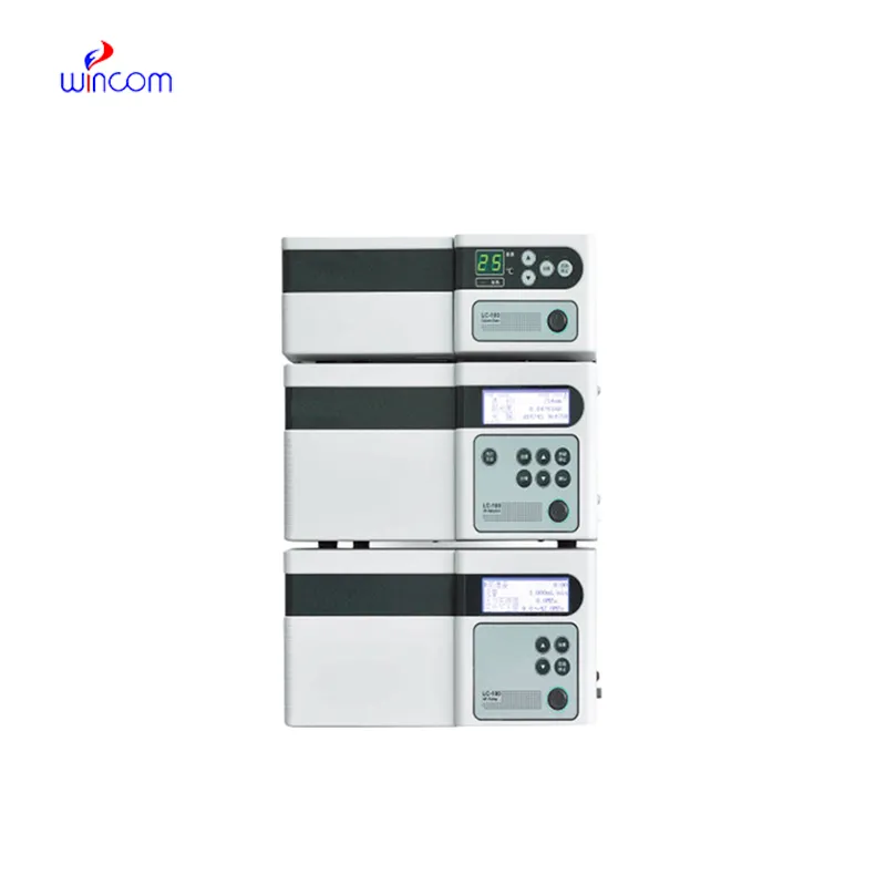
hplc laboratory is a primary tool in hospital and laboratory analytics. Its skills of isolating, measuring, and characterizing both chemical and biological substances enhance research as well as clinical testing. Quality control, drug testing, and testing of samples are done by laboratory technicians using hplc laboratory. The device's flexibility and reliability guarantee uniform performance, yielding critical analytical data that are vital for patient care, experimental validation, and smooth and fast laboratory operations in both healthcare and scientific domains.
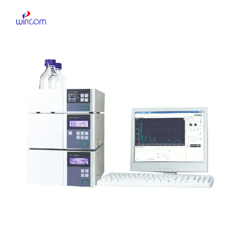
In hplc laboratory used to analyze metabolic profiles and biomarkers during clinical research laboratories. It enables the identification of disease markers and monitoring of biochemical changes over time through the separation of small molecules and proteins. hplc laboratory also facilitates the study of drug absorption and distribution, toxicity testing, and hospital-based clinical trials and thus making it possible to monitor patient responses to therapies in great detail while at the same time ensuring the accuracy and reliability of the analytical results.
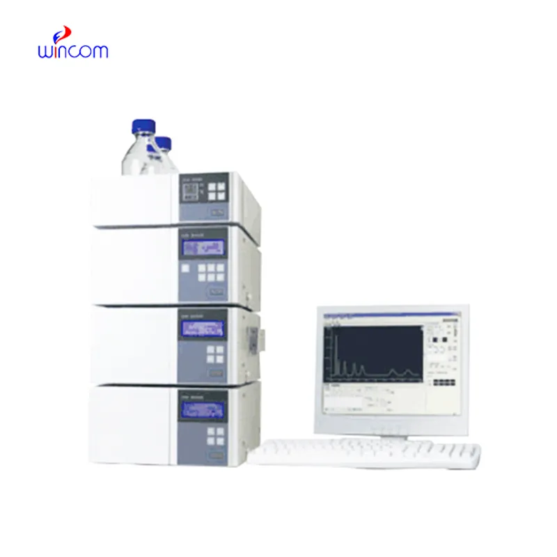
In hplc laboratory, the evolution is probably going to be through miniaturization and portability hplc laboratory is the main feature of the future hospital and laboratory. These advancements will let bedside or point-of-care analysis, thus, improving hospital diagnostics and reducing turnaround times. The future highlights quickness, highly reproducible measurements, and still good accuracy in patient monitoring and laboratory research.
Proper handling and care of hplc laboratory ensure continuous accuracy in the medical laboratory workflows. Cleaning of flow paths, checking detector response, and verifying pump performance are the essential maintenance tasks. Along with the column storage, solvent selection, and routine calibration, laboratory personnel must adhere to the manufacturer guidelines. Proper care enhances reproducibility, reduces downtime, and supports the consistent performance of the laboratory in hospitals and clinical research facilities.
Clinical laboratories make use of hplc laboratory to analyze patient samples with remarkable accuracy. It identifies biomarkers, metabolites, and the levels of therapeutic drugs, thus giving reliable information about the disease status and monitoring treatment. Sensitivity of the technique permits determination of compounds in very minute amounts, which is critical in clinical testing. By resolving complex composition, hplc laboratory guarantees accurate and reproducible results for laboratory diagnostics. Lab staff utilizes it for daily testing, quality control, and research activities, thus making hplc laboratory a vital part of contemporary clinical laboratory work that caters to patient care, treatment choices, and lab data integrity.
Q: What is HPLC used for in laboratories? A: HPLC turns out to be one of the most significant and essential analytical methods in laboratories equipped with the chemical compound analysis, separation, identification, and quantification of their presence in complex samples which are the research, clinical, and pharmaceutical applications. Q: How does HPLC separate compounds? A: The HPLC separation technique is based on the different affinities of the compounds to the stationary phase and mobile phase within the chromatography column. Q: Can HPLC analyze biological samples? A: Yes, it is certainly possible to carry out analyses on various biological fluids such as blood, serum, urine, etc. for the detection of metabolites, drugs, and biomarkers. Q: How often should HPLC columns be replaced? A: The replacement of the columns must be done according to the manufacturer instructions or when the performance begins to decline, which is quite usual after heavy use or contamination. Q: What detectors can be used with HPLC? A: The analysis type determines the use of, among others, UV, fluorescence, refractive index, and mass spectrometry detectors as the common detectors.
We’ve been using this mri machine for several months, and the image clarity is excellent. It’s reliable and easy for our team to operate.
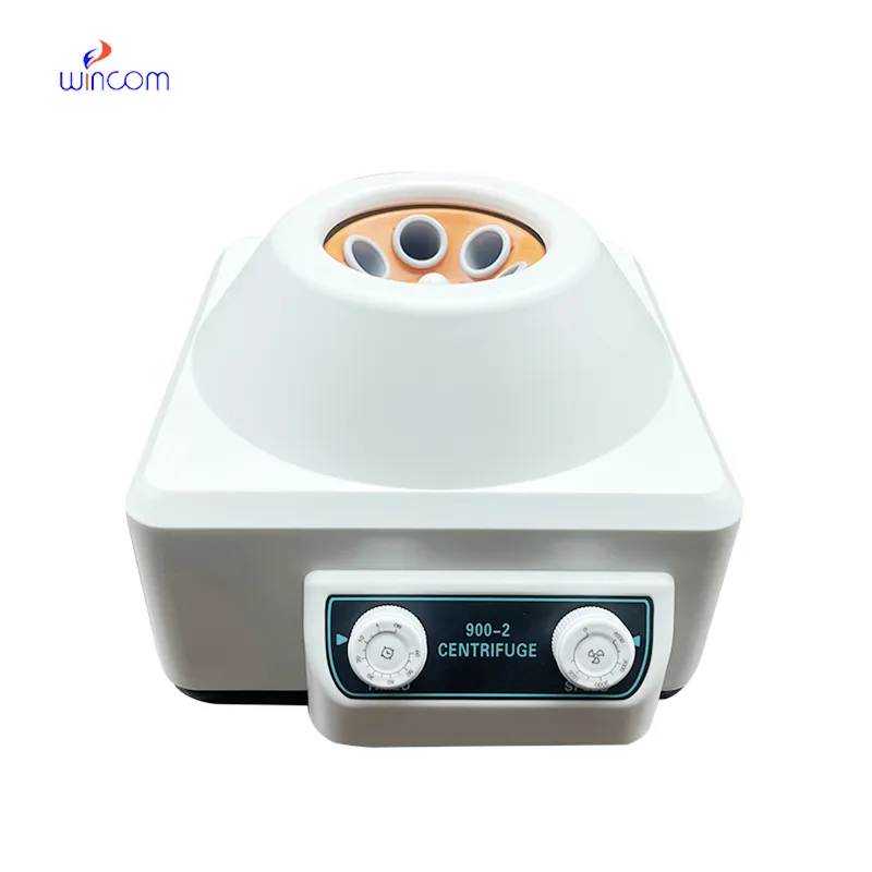
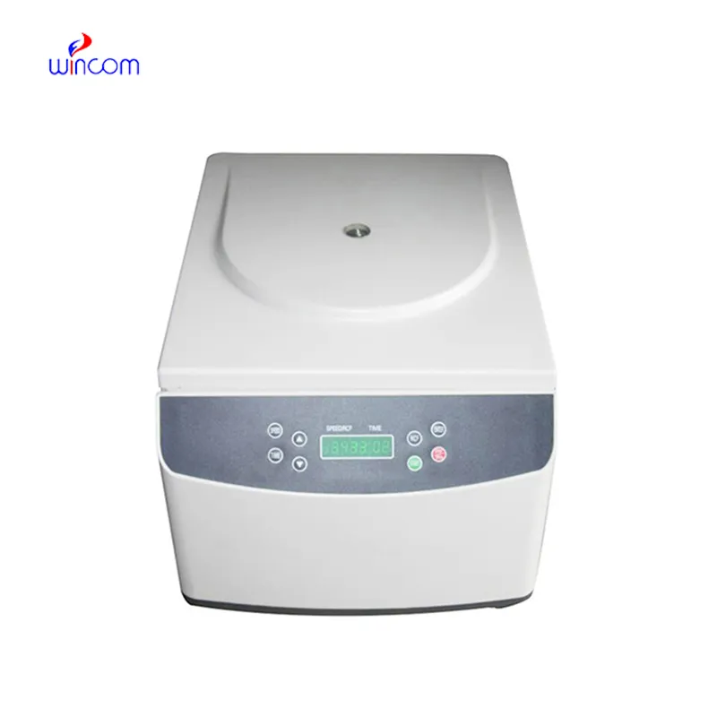
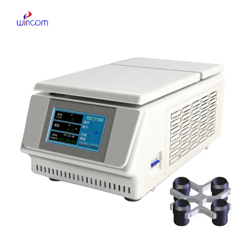
The centrifuge operates quietly and efficiently. It’s compact but surprisingly powerful, making it perfect for daily lab use.
To protect the privacy of our buyers, only public service email domains like Gmail, Yahoo, and MSN will be displayed. Additionally, only a limited portion of the inquiry content will be shown.
I’d like to inquire about your x-ray machine models. Could you provide the technical datasheet, wa...
I’m looking to purchase several microscopes for a research lab. Please let me know the price list ...
E-mail: [email protected]
Tel: +86-731-84176622
+86-731-84136655
Address: Rm.1507,Xinsancheng Plaza. No.58, Renmin Road(E),Changsha,Hunan,China

 af
af
 es
es
 ar
ar
 tr
tr
 sw
sw
 pt
pt
 th
th
 ur
ur
 bn
bn
 ne
ne
 vi
vi
 km
km
 lo
lo
 de
de
 ru
ru
 fi
fi
 nl
nl
 fa
fa
 fr
fr
 ko
ko