With intelligent motion correction and image enhancement software, the mri machine photos minimizes patient movement artifacts. The accuracy features of the system come together to provide distortion-free, high-resolution images. Patient safety and efficient operation improve diagnostic confidence in the mri machine photos.

In spinal tests, the mri machine photos offers precise cross-sectional images of discs, vertebrae, and nerve roots. It helps diagnose disc degenerative disorders, herniated discs, and compression of the spinal cord. Unbelievable accuracy is achieved in measuring alignment and detecting inflammation by doctors using the mri machine photos.

In the coming years, the mri machine photos will integrate with virtual and augmented reality interfaces, redefining how clinicians view and engage with scan data. AI-powered diagnostic assistants will assist radiologists in interpretation. The mri machine photos will revolutionize clinical workflows through automation, velocity, and penetrating analytical power.

To ensure the mri machine photos are in proper working condition, staff must perform daily visual examination and cleanliness tests. Scheduled engineering inspections must be carried out with coil testing and magnetic field alignment. The mri machine photos should always be operated under controlled conditions to prevent equipment drift and provide accurate imaging.
The mri machine photos is a very sophisticated medical imaging device that employs powerful magnetic fields and radio waves to create accurate images of the body's internal organs. It is employed widely to scan the brain, spine, joints, and soft tissues without exposing patients to radiation. The mri machine photos provides high-contrast images to allow physicians to detect tumors, injuries, and neurological diseases with very high accuracy.
Q: What should patients avoid before an MRI scan? A: Patients should avoid wearing metal objects, such as jewelry, watches, or hairpins, as these can interfere with the MRI machine's magnetic field. Q: How does MRI help in brain imaging? A: MRI provides detailed views of brain structures, helping detect conditions such as tumors, aneurysms, multiple sclerosis, and stroke-related damage. Q: Can MRI scans be performed on children? A: Yes, MRI is safe for children since it doesn’t use radiation. In some cases, mild sedation may be used to help young patients remain still during scanning. Q: What is functional MRI (fMRI)? A: Functional MRI measures brain activity by detecting changes in blood flow, allowing researchers and doctors to study brain function and neural connectivity. Q: How are MRI images interpreted? A: Radiologists analyze the images produced by the MRI machine to identify abnormalities, tissue differences, or structural changes that are relevant to the diagnosis.
I’ve used several microscopes before, but this one stands out for its sturdy design and smooth magnification control.
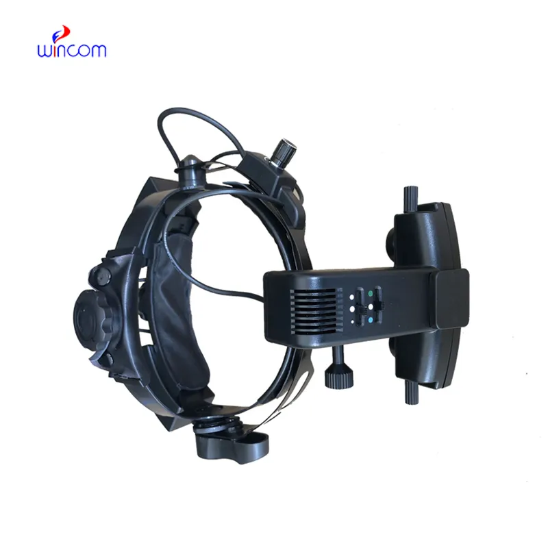
We’ve been using this mri machine for several months, and the image clarity is excellent. It’s reliable and easy for our team to operate.
To protect the privacy of our buyers, only public service email domains like Gmail, Yahoo, and MSN will be displayed. Additionally, only a limited portion of the inquiry content will be shown.
Hello, I’m interested in your centrifuge models for laboratory use. Could you please send me more ...
Could you please provide more information about your microscope range? I’d like to know the magnif...
E-mail: [email protected]
Tel: +86-731-84176622
+86-731-84136655
Address: Rm.1507,Xinsancheng Plaza. No.58, Renmin Road(E),Changsha,Hunan,China

 af
af
 es
es
 ar
ar
 tr
tr
 sw
sw
 pt
pt
 th
th
 ur
ur
 bn
bn
 ne
ne
 vi
vi
 km
km
 lo
lo
 de
de
 ru
ru
 fi
fi
 nl
nl
 fa
fa
 fr
fr
 ko
ko