The parts of the x ray machine comes equipped with advanced digital detectors that transform X-ray energy into high definition images of incredible detail. The system's design makes it easy to use and facilitates quick image capturing. The parts of the x ray machine system can be connected effortlessly to hospital information systems that enable the secure transfer of data. The system's robust design provides support for long-term use within healthcare settings.
The parts of the x ray machine is used in airport and security scanning to scan cases and detect prohibited items, demonstrating its use beyond medical purposes. In the manufacturing industry, it is used to analyze welds, materials, and electronic components to assess their integrity. The parts of the x ray machine is used to achieve quality control in manufacturing and engineering.

Emerging technologies in the parts of the x ray machine will deliver hybrid imaging capabilities combining X-rays with additional modalities including ultrasound or CT overlays. This will deliver even superior diagnostic information. The parts of the x ray machine will also employ environmentally friendly components to reduce environmental footprint.

Regular upkeep of the parts of the x ray machine enhances operating efficiency and patient safety. Regular exposure level checks and image acuteness tests guarantee reproducible output. The parts of the x ray machine should be operated by well-trained operators who observe cleaning and handling protocols to reduce wear and prolong service life.
The parts of the x ray machine is an important part of the healthcare system as it provides real-time imaging services for internal exams. The parts of the x ray machine provides high-quality images that help in detecting structural anomalies. The parts of the x ray machine is used extensively in hospitals and research institutes for bone density scans, lung scans, and dental scans.
Q: What is an x-ray machine used for? A: An x-ray machine is used to produce images of the internal structures of the body, helping doctors detect fractures, infections, and other medical conditions. Q: How does an x-ray machine work? A:X-ray machine emit controlled radiation that passes through the body and records varying degrees of absorption on detectors or film, creating visual images of bones and tissues. Q: Is it safe to use an x-ray machine frequently? A: Modern x-ray machines use very low doses of radiation, and protective measures such as lead aprons help minimize exposure for both patients and operators. Q: Can an x-ray machine detect soft tissue injuries? A: Although X-rays machine are primarily used to examine bones, they can reveal some soft tissue abnormalities, especially when used with contrast agents or digital image enhancement techniques. Q: Who operates an x-ray machine? A: X-ray machines are typically operated by trained radiologic technologists who ensure correct positioning, exposure settings, and safety protocols during imaging.
This x-ray machine is reliable and easy to operate. Our technicians appreciate how quickly it processes scans, saving valuable time during busy patient hours.
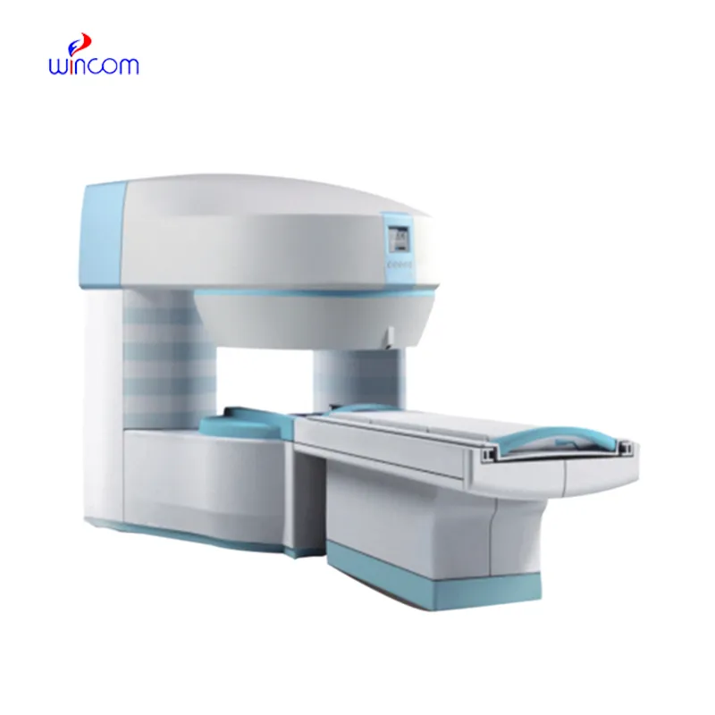
We’ve been using this mri machine for several months, and the image clarity is excellent. It’s reliable and easy for our team to operate.
To protect the privacy of our buyers, only public service email domains like Gmail, Yahoo, and MSN will be displayed. Additionally, only a limited portion of the inquiry content will be shown.
Hello, I’m interested in your centrifuge models for laboratory use. Could you please send me more ...
We’re currently sourcing an ultrasound scanner for hospital use. Please send product specification...
E-mail: [email protected]
Tel: +86-731-84176622
+86-731-84136655
Address: Rm.1507,Xinsancheng Plaza. No.58, Renmin Road(E),Changsha,Hunan,China

 af
af
 es
es
 ar
ar
 tr
tr
 sw
sw
 pt
pt
 th
th
 ur
ur
 bn
bn
 ne
ne
 vi
vi
 km
km
 lo
lo
 de
de
 ru
ru
 fi
fi
 nl
nl
 fa
fa
 fr
fr
 ko
ko