The shimadzu x ray machine comes equipped with an intelligent imaging system that increases grayscale depth and detail understanding. The complex algorithms of the shimadzu x ray machine improve the viewing of subtle lesions and tissues. The shimadzu x ray machine has been designed for high throughput capabilities that promote rapid viewing cycles and convenient data accessibility.
The shimadzu x ray machine is used extensively in dentistry, yielding minute-level images of teeth, jaw bones, and surrounding tissues. It assists in the diagnosis of cavities, orthodontic problems, and impacted tooth diagnosis. The shimadzu x ray machine is used for endodontic and implant planning to allow precision treatment delivery.
The shimadzu x ray machine of the future will target integrating artificial intelligence to aid image interpretation and identify anomalies. Analysis software will automatically detect early-stage diseases more accurately. The shimadzu x ray machine will further feature low-dose radiation technologies, which will ensure that imaging is safer, more sustainable for both patients and operators.
The life of the shimadzu x ray machine relies on proper maintenance and surveillance. The X-ray tube, generator, and control panel are some of the parts that need to be examined and serviced based on manufacturer recommendations. The shimadzu x ray machine should be protected from moisture, vibration, and heavy dust to prevent performance loss.
The shimadzu x ray machine is intended to generate high-quality radiographic images that can capture internal details exceptionally. The device has the capability of being used in several medical functions such as trauma analysis and disease diagnosis. The portable and efficient shimadzu x ray machine helps in speeding up diagnosis and providing better treatment to the patient.
Q: What makes an x-ray machine different from a CT scanner? A: An x-ray machine captures a single 2D image, while a CT scanner takes multiple x-rays from different angles to create 3D cross-sectional views. Q: How is image quality measured in an x-ray machine? A: Image quality depends on factors like contrast, resolution, and exposure settings, which are adjusted based on the target area being examined. Q: What power supply does an x-ray machine require? A: Most x-ray machines operate on high-voltage power systems, typically between 40 to 150 kilovolts, depending on their intended use. Q: Can x-ray machines be used for dental imaging? A: Yes, specialized dental x-ray machines provide detailed images of teeth, jaws, and surrounding structures to support oral health assessments. Q: How does digital imaging improve x-ray efficiency? A: Digital systems allow instant image preview, faster diagnosis, and reduced need for retakes, improving workflow efficiency in clinical environments.
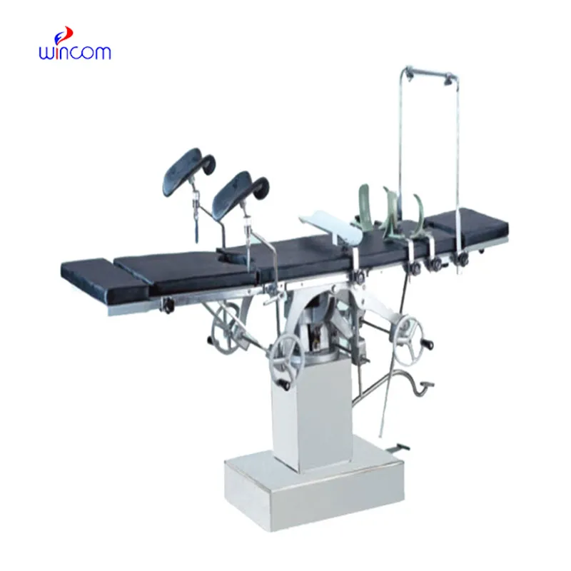
The delivery bed is well-designed and reliable. Our staff finds it simple to operate, and patients feel comfortable using it.
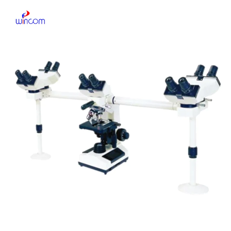
The microscope delivers incredibly sharp images and precise focusing. It’s perfect for both professional lab work and educational use.
To protect the privacy of our buyers, only public service email domains like Gmail, Yahoo, and MSN will be displayed. Additionally, only a limited portion of the inquiry content will be shown.
I’d like to inquire about your x-ray machine models. Could you provide the technical datasheet, wa...
I’m looking to purchase several microscopes for a research lab. Please let me know the price list ...
E-mail: [email protected]
Tel: +86-731-84176622
+86-731-84136655
Address: Rm.1507,Xinsancheng Plaza. No.58, Renmin Road(E),Changsha,Hunan,China

 af
af
 es
es
 ar
ar
 tr
tr
 sw
sw
 pt
pt
 th
th
 ur
ur
 bn
bn
 ne
ne
 vi
vi
 km
km
 lo
lo
 de
de
 ru
ru
 fi
fi
 nl
nl
 fa
fa
 fr
fr
 ko
ko